


ISSN 1757-8515
Predictors Of Difficult Intubation: Study In Kashmiri Population
Arun Kr. Gupta, Mohamad Ommid, Showkat Nengroo, Imtiyaz Naqash and Anjali Mehta
Cite this article as: BJMP 2010;3(1):307
|
|
Abstract Airway assessment is the most important aspect of anaesthetic practice as a difficult intubation may be unanticipated. A prospective study was done to compare the efficacy of airway parameters to predict difficult intubation. Parameters studied were degree of head extension, thyromental distance, inter incisor gap, grading of prognathism, obesity and modified mallampati classification. 600 Patients with ASA I& ASA II grade were enrolled in the study. All patients were preoperatively assessed for airway parameters. Intra-operatively all patients were classified according to Cormack and Lehane laryngoscopic view. Clinical data of each test was collected, tabulated and analyzed to obtain the sensitivity, specificity, positive predictive value & negative predictive value. Results obtained showed an incidence of difficult intubation of 3.3 % of patients. Head and neck movements had the highest sensitivity (86.36%); high arched palate had the highest specificity (99.38%). Head and neck movements strongly correlated for patients with difficult intubation. Keywords: Intubation, Anaesthesia, Laryngoscopy |
Introduction
The fundamental responsibility of an anesthesiologist is to maintain adequate gas exchange through a patent airway. Failure to maintain a patent airway for more than a few minutes results in brain damage or death1. Anaesthesia in a patient with a difficult airway can lead to both direct airway trauma and morbidity from hypoxia and hypercarbia. Direct airway trauma occurs because the management of the difficult airway often involves the application of more physical force to the patient’s airway than is normally used. Much of the morbidity specifically attributable to managing a difficult airway comes from an interruption of gas exchange (hypoxia and hypercapnia), which may then cause brain damage and cardiovascular activation or depression2.
Though endotracheal intubation is a routine procedure for all anesthesiologists, occasions may arise when even an experienced anesthesiologist might have great difficulty in the technique of intubation for successful control of the airway. As difficult intubation occurs infrequently and is not easy to define, research has been directed at predicting difficult laryngoscopy, i.e. when is not possible to visualize any portion of the vocal cords after multiple attempts at conventional laryngoscopy. It is argued that if difficult laryngoscopy has been predicted and intubation is essential, skilled assistance and specialist equipment should be provided. Although the incidence of difficult or failed tracheal intubation is comparatively low, unexpected difficulties and poorly managed situations may result in a life threatening condition or even death3.
Difficulty in intubation is usually associated with difficulty in exposing the glottis by direct laryngoscopy. This involves a series of manoeuvres, including extending the head, opening the mouth, displacing and compressing the tongue into the submandibular space and lifting the mandible forward. The ease or difficulty in performing each of these manoeuvres can be assessed by one or more parameters4.
Extension of the head at the atlanto-occipital joint can be assessed by simply looking at the movements of the head, measuring the sternomental distance, or by using devices to measure the angle5. Mouth opening can be assessed by measuring the distance between upper and lower incisors with the mouth fully open. The ease of lifting the mandible can be assessed by comparing the relative position of the lower incisors in comparison with the upper incisors after forward protrusion of the mandible6. The measurement of the mento-hyoid distance and thyromental distance provide a rough estimate of the submandibular space7. The ability of the patient to move the lower incisor in front of the upper incisor tells us about jaw movement. The classification provided by Mallampati et al8 and later modified by Samsoon and Young9 helps to assess the size of tongue relative to the oropharynx. Abnormalities in one or more of these parameters may help predict difficulty in direct laryngoscopy1.
Initial studies attempted to compare individual parameters to predict difficult intubation with mixed results8,9. Later studies have attempted to create a scoring system3,10 or a complex mathematical model11,12. This study is an attempt to verify which of these factors are significantly associated with difficult exposure of glottis and to rank them according to the strength of association.
Materials & methods
The study was conducted after obtaining institutional review board approval. Six hundred ASA I & II adult patients, scheduled for various elective procedures under general anesthesia, were included in the study after obtaining informed consent. Patients with gross abnormalities of the airway were excluded from the study. All patients were assessed the evening before surgery by a single observer. The details of airway assessment are given in Table I.
Table I: Method of assessment of various airway parameters (predictors)
|
Airway Parameter
|
Method of assessment
|
|
Modified Mallampati Scoring
|
Class I: Faucial pillars, soft palate and uvula visible.
Class II: Soft palate and base of uvula seen
Class III: Only soft palate visible.
Class IV: Soft palate not seen
Class I & II : Easy Intubation
Class III & IV: Difficult Intubation
|
|
Obesity
|
Obese BMI (≥ 25)
Non Obese BMI (< 25)
|
|
Inter Incisor Gap
|
Distance between the incisors with mouth fully open(cms)
|
|
Thyromental distance
|
Distance between the tip of thyroid cartilage and tip of chin, with fully extended(cms)
|
|
Degree of Head Extension
|
Grade I ≥ 90◦
Grade II = 80◦-90◦
Grade III < 80◦
|
|
Grading of Prognathism
|
Class A: - Lower incisor protruded anterior to the upper incisor.
Class B: - Lower incisor brought edge to edge with upper incisor but not anterior to them.
Class C: - Lower incisors could be brought edge to edge.
|
In addition the patients were examined for the following.
- High arched palate.
- Protruding maximally incisor (Buck teeth)
- Wide & short Neck
Direct laryngoscopy with Macintosh blade was performed by an anaesthetist who was blinded to preoperative assessment.
Glottic exposure was graded as per Cormack-Lehane classification13 (Fig 1).
Figure 1: Cormack-Lehane grading of glottic exposure on direct laryngoscopy
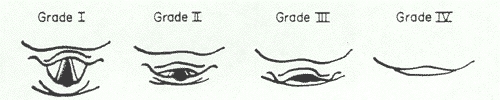
Grade 1: most of the glottis visible; Grade 2: only the posterior extremity of the glottis and the epiglottis visible; Grade 3: no part of the glottis visible, only the epiglottis seen; Grade 4: not even the epiglottis seen. Grades 1 and 2 were considered as ‘easy’ and grades 3 and 4 as ‘difficult’.
Results
Glottic exposure on direct laryngoscopy was difficult in 20 (3.3%) patients.
The frequency of patients in various categories of ‘predictor’ variables is given in Table-II
Table II: The frequency analysis of predictor parameters
|
Airway Parameter
|
Group
|
Frequency (%)
|
|
Modified Mallampati Scoring
|
Class 1&2
Class 3&4
|
96%
4%
|
|
Obesity
|
Obese BMI (≥ 25)
Non Obese BMI (< 25)
|
28.7%
71.3%
|
|
Inter Incisor Gap
|
Class I : >4cm
Class II: <4cm
|
93.5%
6.5%
|
|
Thyromental distance
|
Class I: ≥ 6cm.
Class II: ≤6cm.
|
94.6%
5.4%
|
|
Head & Neck Movements
|
Difficult {class II & III (90˚)}
Easy {class I(>90˚)}
|
16%
84%
|
|
Grading of Prognathism
|
Difficult (class III)
Easy (class I + II)
|
96.1%
3.9%
|
|
Wide and Short neck
|
Normal neck body ratio 1:13
Difficult (Ratio≥ 1:13)
|
86.9%
13.1%
|
|
High arched Palate
|
Yes
No
|
1.9%
98.1%
|
|
Protruding Incisors
|
Yes
No
|
4.2%
95.8%
|
The association between different variables and difficulty in intubation was evaluated using the chi-square test for qualitative data and the student’s test for quantitative data and p<0.05 was regarded as significant. The clinical data of each test was used to obtain the sensitivity, specificity and positive and negative predictive values. Results are shown in Table III.
Table III: Comparative analysis of various physical factors and scoring systems
|
Physical factors and various Scoring Systems
|
Sensitivity ( % )
|
Specificity ( % )
|
PPV
( % )
|
NPV
( % )
|
|
Obesity
|
81.8
|
72.76
|
6.34
|
99.43
|
|
Inter incisor gap
|
18.8
|
94.14
|
6.6
|
98.1
|
|
Thyromental distance
|
72.7
|
96.5
|
32.0
|
99.4
|
|
Head and Neck movement
|
86.36
|
86.0
|
34.6
|
99.7
|
|
Prognathism
|
4.5
|
96.3
|
2.7
|
97.9
|
|
Wide and Short neck
|
45.5
|
87.9
|
7.8
|
98.6
|
|
High arched palate
|
40.1
|
99.38
|
60.0
|
98.67
|
|
Protruding incisor
|
4.6
|
95.9
|
2.5
|
97.79
|
|
Mallampati scoring system
|
77.3
|
98.2
|
48.57
|
99.5
|
|
Cormack and Lehane’s scoring system
|
100
|
99.7
|
88
|
100
|
Discussion
Difficulty in endotracheal intubation constitutes an important cause of morbidity and mortality, especially when it is not anticipated preoperatively. This unexpected difficulty in intubation is the result of a lack of accurate predictive tests and inadequate preoperative assessment of the airway. Risk factors if identified at the preoperative visit help to alert the anaesthetist so that alternative methods of securing the airway can be used or additional expertise sought before hand.
Direct laryngoscopy is the gold standard for tracheal intubation. There is no single definition of difficult intubation but the ASA defines it as occurring when “tracheal intubation requires multiple attempts, in the presence or absence of tracheal pathology”. Difficult glottic view on direct laryngoscopy is the most common cause of difficult intubation. The incidence of difficult intubation in this study is similar to that found in others.
As for as the predictors are concerned, different parameters for the prediction of difficult airways have been studied. Restriction of head and neck movement and decreased mandibular space have been identified as important predictors in other studies. Mallampati classification has been reported to be a good predictor by many but found to be of limited value by others14. Interincisor gap, forward movement of jaw and thyromental distance have produced variable results in predicting difficult airways in previous studies7,15. Even though thyromental distance is a measure of mandibular space, it is influenced by degree of head extension.
There have been attempts to create various scores in the past. Many of them could not be reproduced by others or were shown to be of limited practical value. Complicated mathematical models based on clinical and/or radiological parameters have been proposed in the past16, but these are difficult to understand and follow in clinical settings. Many of these studies consider all the parameters to be of equal importance.
Instead of trying to find ‘ideal’ predictor(s), scores or models, we simply arranged them in an order based on the strength of association with difficult intubation. Restricted extension of head, decreased thyromental distance and poor Mallampati class are significantly associated with difficult intubation.
In other words patients with decreased head extension are at much higher risk of having a difficult intubation compared to those with abnormalities in other parameters. The type of equipment needed can be chosen according to the parameter which is abnormal. For example in a patient with decreased mandibular space, it may be prudent to choose devices which do not involve displacement of the tongue like the Bullard laryngoscope or Fiber-optic laryngoscope. Similarly in patients with decreased head extension devices like the McCoy Larngoscope are likely to be more successful.
Conclusion
This prospective study assessed the efficacy of various parameters of airway assessment as predictors of difficult intubation. We have find that head and neck movements, high arched palate, thyromental distance & Modified Malampatti classification are the best predictors of difficult intubation.
This prospective study assessed the efficacy of various parameters of airway assessment as predictors of difficult intubation. We have find that head and neck movements, high arched palate, thyromental distance & Modified Malampatti classification are the best predictors of difficult intubation.
|
Competing Interests None Declared Author Details ARUN KUMAR GUPTA, Dept. Of Anaesthesiology, Rural Medical College, Loni, MOHAMED OMMID, Dept. Of Anaesthesiology, SKIMS, Soura, J&K, India SHOWKAT NENGROO, Dept. Of Anaesthesiology, SKIMS, Soura, J&K,India IMTIYAZ NAQASH, Dept. Of Anaesthesiology, SKIMS, Soura, J&K,India ANJALI MEHTA, Dept. Of Anaesthesiology, GMC Jammu, J&K, India CORRESPONDENCE: ARUN KUMAR GUPTA, Assistant Professor Dept. of Anaesthesiology & Critical Care Rural Medical College, Loni Maharashtra, India, 413736 Email: guptaarun71@yahoo.com |
References
1.Rose DK, Cohen MM. The airway: problems and predictions in 18,500 patients. Can J Anaesth 1994; 41:372-83.
2.Benumof JL: Management of the difficult airway: With special emphasis on awake tracheal intubation. Anesthesiology 1991; 75: 1087-1110
3.Wilson ME, Spiegelhalter D, Robertson JA, Lesser P. Predicting difficult intubation. Br J Anaesth 1988; 61(2):211-6.
4. A.Vasudevan, A.S.Badhe. Predictors of difficult intubation – a simple approach. The Internet Journal of Anesthesiology2009; 20(2)
5.Tse JC, Rimm EB, Hussain A. Predicting difficult endotracheal intubation in surgical patients scheduled for general anesthesia: a prospective blind study. Anesth Analg 1995; 81(2):254-8.
6.Savva D. Prediction of difficult tracheal intubation. Br J Anaesth 1994; 73(2):149-53.
7.Butler PJ, Dhara SS. Prediction of difficult laryngoscopy: an assessment of the thyromental distance and Mallampati predictive tests. Anaesth Intensive Care 1992; 20(2):139-42.
8.Mallampati SR, Gatt SP, Gugino LD, Desai SP, Waraksa B, Freiberger D, et al. A clinical sign to predict difficult tracheal intubation: a prospective study. Can Anaesth Soc J 1985; 32(4):429-34.
9.Samsoon GL, Young JR. Difficult tracheal intubation: a retrospective study. Anaesthesia 1987; 42(5):487-90.
10. Nath G, Sekar M. Predicting difficult intubation--a comprehensive scoring system. Anaesth Intensive Care 1997; 25(5):482-6.
11. Charters P. Analysis of mathematical model for osseous factors in difficult intubation. Can J Anaesth 1994; 41(7):594-602.
12. Arne J, Descoins P, Fusciardi J, Ingrand P, Ferrier B, Boudigues D, et al. Preoperative assessment for difficult intubation in general and ENT surgery: predictive value of a clinical multivariate risk index. Br J Anaesth 1998; 80(2):140-6.
13. Cormack RS, Lehane J. Difficult tracheal intubation in obstetrics. Anaesthesia 1984; 39(11):1105-11.
14. Lee A, Fan LT, Gin T, Karmakar MK, Ngan Kee WD. A systematic review (meta-analysis) of the accuracy of the Mallampati tests to predict the difficult airway. Anesth Analg 2006; 102(6):1867-78.
15. Bilgin H, Ozyurt G. Screening tests for predicting difficult intubation. A clinical assessment in Turkish patients. Anaesth Intensive Care 1998; 26(4):382-6.
16. Naguib M, Malabarey T, AlSatli RA, Al Damegh S, Samarkandi AH. Predictive models for difficult laryngoscopy and intubation. A clinical, radiologic and three-dimensional computer imaging study. Can J Anaesth 1999; 46(8):748-59.

The above article is licensed under a Creative Commons Attribution-NonCommercial-NoDerivatives 4.0 International License.