Patellar fracture fixation: An unreported complication occurring directly attributable to tension band fixation of the patella.
Peter Alexander Gilmer Torrie, James, Smith and Micheal Kelly.
Cite this article as: BJMP 2014;7(1):a703
|
|
Abstract Patella fractures account for 1% of all fractures but there is little in the contemporary literature regarding either an optimal standardized post-operative rehabilitation regimen or the long-term outcomes following these fractures. Tension band wire fixation for displaced patella fractures is a well-recognised and accepted method of operative treatment for these fractures. In this case the authors report a new complication, yet to be documented in the literature that was directly attributable to a well-recognised complication resulting from this method of fixation. An atypical osteochondral defect, from the lateral femoral condyle, was generated as a direct result of bony spur at the site of the previous patella fracture malunion. As the patient fell on to her knee the bony spur was driven into the femoral condyle in a similar fashion to an osteotome, generating the atypical osteochondral defect. This patient had chronic anterior knee pain following the tension band fixation of her patella. An inadequate standardised follow-up regimen failed to identify her fracture malunion that was responsible for her ongoing persistent symptoms. Only as a result of this previously unreported complication, we were able to identify and surgically address the underlying primary pathology responsible for her persistent symptoms. This case highlights the importance for the identification and establishment of a more robust imaging follow-up regimen post patella fracture fixation. Keywords: patella fracture; tension band fixation; unreported complication; post-operative follow-up regimen |
Introduction
Patella fractures account for 1% of all fractures but there is little in the contemporary literature regarding outcomes. It is accepted that where fixation is required it needs to be rigid and tension band wiring using cannulated screws or k-wires is the accepted standard.1 Recognised complications associated with this form of fixation occur in up to 15% of cases2 and include; infection, loss of fixation, knee stiffness, post-traumatic osteoarthritis, malunion, nonunion and irritation from hardware.3Thereis nothing in the literature regarding the natural history following fixation.
We report an unusual complication of an osteochondral defect being generated as a direct result of a malunion of a patella fracture previously fixed by a tension band wiring technique.
Case Report
A 35-year old lady presented to our unit after a direct fall on to her left knee with an associated dislocation of her patella that spontaneously reduced on extension, at the time of injury. Three years previously she had sustained a patella fracture that had been treated with tension band wiring. From the time of the original fixation she had experienced mild persistent anterior knee pain, with a reduced range of motion and grinding. She had been discharged from further follow up.
On this presentation, examination revealed that she had a marked knee effusion with a functional extensor mechanism and a range of motion from 0-60 degrees.
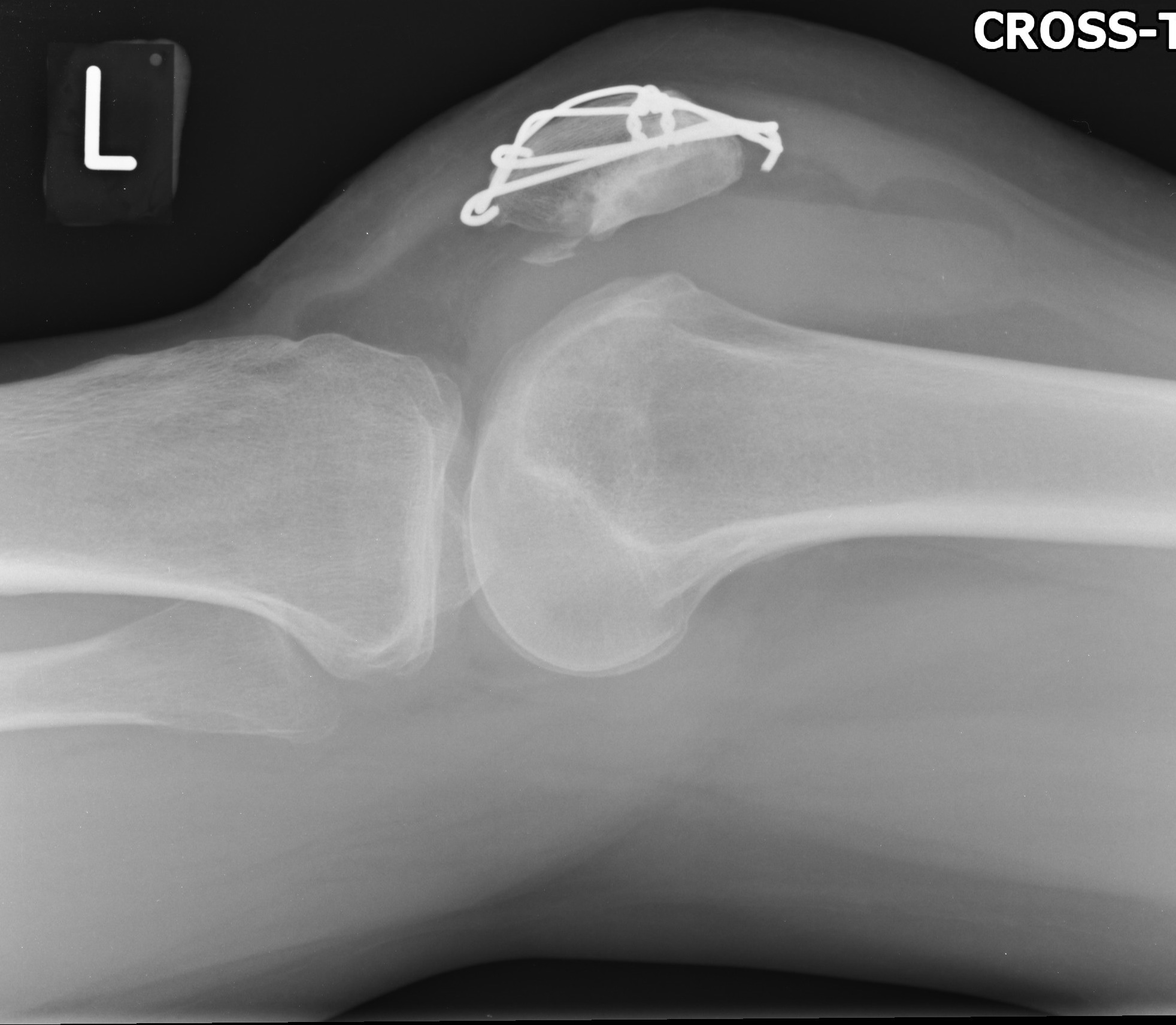
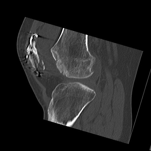
The initial radiographs demonstrated that she had broken hardware and an incongruency of the patella suggesting malunion on the articular surface with a residual step (figure 1). In addition, an osteochondral fragment was identified in the patellofemoral joint. Computer tomography was undertaken and confirmed the osteochondral fragment had come from the lateral femoral condyle and a spur like prominence on the articular surface of the patella (figure 2). The mechanism was that this bony spur had been driven into the articular surface of the lateral condyle on dislocation resulting in the osteochondral fragment being generated.
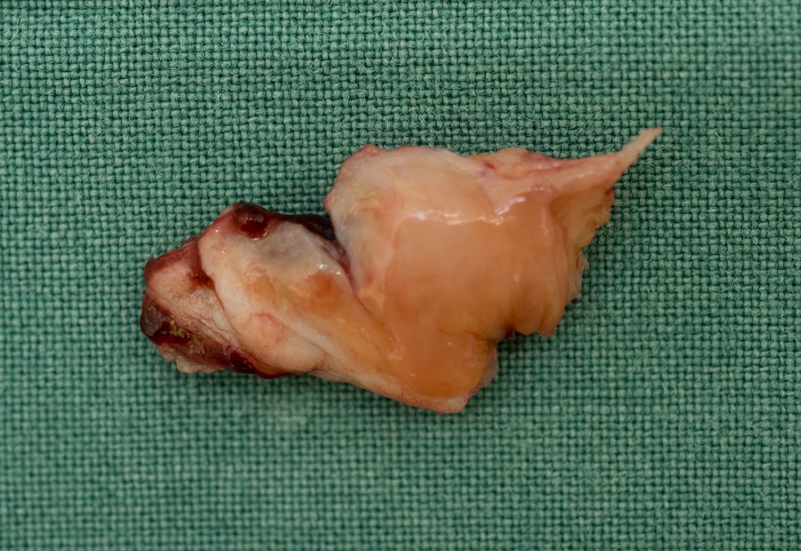
Intraoperative findings confirmed this and measured the osteochondral fragment as 40x15mm (figure 3). In addition it was found that the lateral longitudinal wire had protruded into the joint causing a vertical linear defect in the articular surface of the trochlea.
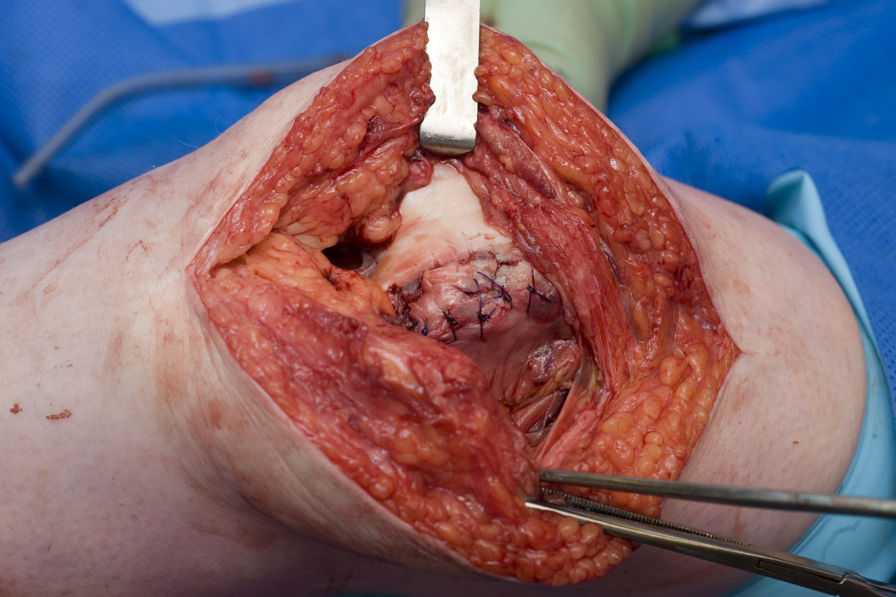
The osteochondral defect was reduced and stabilized with interrupted 3/0 PDS sutures achieving a smooth articular surface (figure 4). In addition a patelloplasty and a lateral release were performed to remove the bony prominence and restore patella tracking respectively. At 6-month follow up this patient was progressing well with rehabilitation and the majority of her chronic symptoms had resolved.
Figure 1. Lateral radiograph demonstrating an incongruency of the patella suggesting malunion on the articular surface with a residual step
Figure 2. Computerised Tomography confirming an osteochondral fragment that had come from the lateral femoral condyle. 
Figure 3. Osteochondral fragment from the lateral femoral condyle measuring 40x15mm.
Figure 4. The osteochondral defect stabilized with interrupted 3/0 PDS sutures achieving a smooth articular surface
Discussion
Although patellae account for 1% of fractures their functional outcome remains largely ignored in the literature. This case presents an unreported complication and highlights that symptoms can remain following the initial fixation that are accepted either by the patient or the treating centre and not further investigated.
Osteoarthritis of the knee remains the most common musculoskeletal complaint in general practice but even then only a third of those with symptoms seek medical advice. Therefore the lack of re-referrals following fixation is not an accurate way to assess outcome following patella fractures treated with this mode of fixation.
Patella fractures represent only a small number of fractures and therefore assessment of treatment and outcomes is problematic, particularly as there is no standardised rehabilitation regimen.
We report on this case as it illustrates a complication of patella fracture fixation that has not been previously described or routinely recognised and, additionally, highlights the fundamental importance of a comprehensive standardised post-operative imaging follow-up regimen. It may be that patients are not currently being correctly counselled regarding the longer-term expectations following patella fracture. A study to define the natural history of patella fractures following contemporary management is needed.
|
Competing Interests None declared Author Details Peter Torrie MB ChB, MRCS (Eng), PGCertEd; James Smith MRCS (Eng); Michael Kelly FRCS (Tr + Orth); Bristol, UK. CORRESPONDENCE: Mr P.A.G Torrie – Flat 12, Muller House, Ashley Down Road, Bristol, BS7 9DA, UK. Email: alextorrie99@hotmail.com |
References
- Smith ST, Cramer KE, Karges DE et al. Early complications in the operative treatment of patellar fractures. J Orthop Trauma. 1997 Apr;11(3):183-7.
- Bostman O, Kiviluoto O, Santavirta S, et al. Fractures of the patella treated by operation. Arch Orthop Trauma Surg. 1983; 102:78-81.
- Carpenter JE, Kasman R, Matthews LS. Fractures of the patella. J Bone Joint Surg Am. 1993;75:1550-1561.

The above article is licensed under a Creative Commons Attribution-NonCommercial-NoDerivatives 4.0 International License.


